
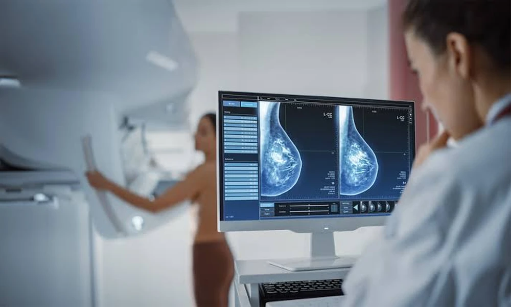
A routine mammogram reveals an area of concern. Your doctor recommends additional imaging. This scenario affects thousands of women each year, and while it’s natural to feel anxious, understanding what comes next can help you approach the situation with clarity rather than fear. The journey from initial screening to diagnosis involves several imaging technologies, each designed to provide specific information about breast tissue.
Why Screening Results Require Follow-Up
Mammography remains the gold standard for breast cancer screening, but it has limitations. Dense breast tissue, which appears white on mammograms just like tumors do, can obscure abnormalities. According to radiological standards, approximately 10% of screening mammograms lead to a callback for additional imaging. The vast majority of these callbacks result in benign findings, but the process exists to catch potential problems early.
When radiologists identify something unusual-a mass, calcifications, or an area of architectural distortion-they need more detailed images. This is where diagnostic mammography, ultrasound, and advanced magnetic resonance imaging enter the picture. Each technology offers different strengths in visualizing breast tissue and determining whether an abnormality requires tissue sampling.
The Role of Ultrasound in Breast Evaluation
Ultrasound uses sound waves to create real-time images of breast tissue. It excels at distinguishing fluid-filled cysts from solid masses, a critical distinction that often immediately resolves concerns. A simple cyst typically requires no further action, while solid masses need characterization.
For women with dense breast tissue, supplemental ultrasound screening can detect cancers that mammography misses. The technology is particularly useful for younger women whose denser tissue composition makes mammography less effective. Ultrasound also guides many breast biopsies, allowing physicians to target suspicious areas with precision while watching the needle placement in real time.
When Magnetic Resonance Imaging Becomes Necessary
Breast MRI provides the most detailed imaging available, using powerful magnets and radio waves to create cross-sectional images. Unlike mammography and ultrasound, MRI can detect abnormalities based on how tissue behaves rather than just how it looks. Cancerous tissue typically shows increased blood flow, which appears as enhancement after contrast injection.
High-risk women-those with BRCA gene mutations, strong family histories, or previous chest radiation-often undergo annual MRI screening alongside mammography. MRI can also assess the extent of known cancer, check for disease in the opposite breast, and evaluate treatment response. However, MRI’s sensitivity comes with a tradeoff: it sometimes flags benign tissue as suspicious, leading to unnecessary biopsies.
Advanced Biopsy Techniques for Accurate Diagnosis
When imaging cannot definitively rule out cancer, tissue sampling provides the answer. Modern biopsy techniques have evolved significantly from surgical excision, which was once the only option. Core needle biopsy, performed with image guidance, retrieves several small tissue samples through a single insertion point. This minimally invasive approach provides sufficient tissue for pathological analysis while avoiding the operating room.
For abnormalities visible only on MRI, specialized facilities offer mri biopsy procedures. This technique allows precise targeting of suspicious areas that other imaging modalities cannot adequately visualize. The procedure takes place inside the MRI scanner, where real-time imaging guides needle placement to the exact location identified on diagnostic scans. While more complex than ultrasound-guided biopsy, this approach ensures that concerning findings don’t go unsampled simply because they’re invisible on conventional imaging.
The pathology results typically arrive within a few days to a week. Benign results mean no cancer is present, though some findings may require monitoring. Malignant results lead to staging studies and treatment planning. Atypical or high-risk findings might warrant surgical removal even without cancer, as these conditions increase future cancer risk.
Managing the Emotional Impact of Uncertain Results
The waiting period between screening and final diagnosis tests anyone’s emotional resilience. Anxiety during this time is completely normal, but excessive worry serves no protective function. Research shows that managing stress during medical uncertainty requires deliberate strategies rather than simply trying to “stay positive.”
Practical stress management includes maintaining routines, limiting repetitive information searches, and acknowledging feelings without letting them dictate behavior. Many people find that structured mindfulness practices help regulate the nervous system during periods of medical uncertainty. The goal is not to eliminate concern but to prevent it from becoming overwhelming.
Some women benefit from bringing a trusted person to appointments, which helps with information retention and emotional support. Writing down questions before appointments ensures you address your concerns rather than thinking of them afterward. Understanding the timeline for each step-from imaging to biopsy to results-provides structure during an inherently uncertain process.
Access to Specialized Diagnostic Services
Geographic location significantly impacts access to advanced diagnostic imaging. While basic mammography and ultrasound are widely available, MRI-guided biopsy requires specialized equipment and trained radiologists. Urban medical centers typically offer the full range of diagnostic services, but patients in smaller communities may need to travel for certain procedures.
International patients increasingly seek specialized medical procedures in facilities with advanced technology and experienced specialists. This trend reflects both the concentration of cutting-edge equipment in certain centers and the importance of radiologist experience in accurately interpreting complex imaging studies.
What Happens After Diagnosis
Benign biopsy results typically lead to a return to routine screening schedules, though some findings warrant shorter surveillance intervals. If cancer is diagnosed, the next phase involves determining its characteristics-hormone receptor status, HER2 status, and grade-which guide treatment decisions.
Modern breast cancer treatment is highly individualized. Stage, tumor biology, patient age, and overall health all factor into the treatment plan. Options range from lumpectomy with radiation for small, early-stage tumors to mastectomy with reconstruction for more extensive disease. Systemic treatments-chemotherapy, hormone therapy, or targeted drugs-depend on tumor characteristics rather than size alone.
The multidisciplinary approach means you’ll likely meet with several specialists: a surgeon, a medical oncologist, and perhaps a radiation oncologist. Each provides expertise in their domain, and together they develop a coordinated treatment strategy. This team-based care has become standard because breast cancer treatment now involves multiple modalities working in sequence or combination.
Moving Forward with Information
The transition from screening to diagnosis involves multiple steps, each designed to provide specific information. Understanding this process helps you participate actively in your care rather than feeling swept along by events. Ask questions when something isn’t clear. Request copies of your imaging reports. Know that additional testing doesn’t mean cancer is present-it means your medical team is being thorough.
Early detection remains one of the most powerful tools in breast cancer management. While the diagnostic process can feel overwhelming, it exists to catch problems when they’re most treatable. Whether your results show benign findings or require treatment, you’ll face the situation with accurate information rather than ambiguity. That clarity, though sometimes hard-won, forms the foundation for whatever comes next.